14++ Cystic Hygroma Ultrasound
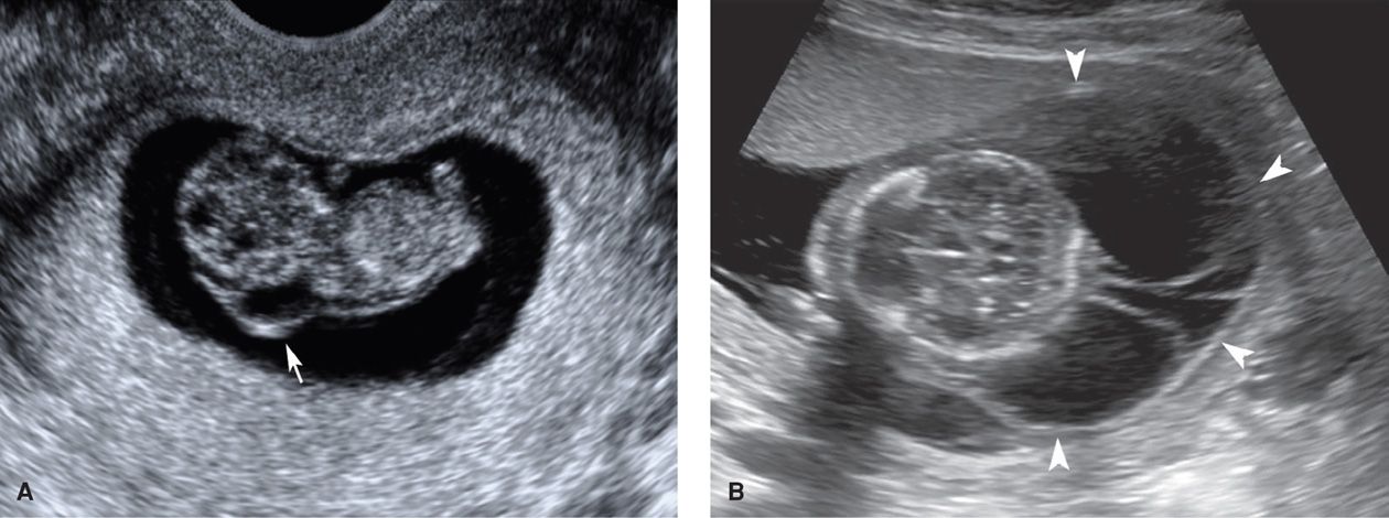
Cystic Hygroma Ultrasound. Cystic hygroma (ch) is a fetal sonographic finding with an incidence of 1%. Cystic hygroma is a rare congenital malformation of the lymphatic system, most frequently detected in the head and neck region.

Reprinted with permission from thefetus.net Cystic hygromas are single or multiple cysts found mostly in the neck region. The prenatal diagnosis of cystic hygroma using ultrasound is well documented in the literature.
ducati x diavel s price in india dalle beton gravillonnee 50x50 brico depot dessin de cheminee de noel facile du papier peint
Evaluation of fetal arrhythmias from simultaneous pulsed
This fluid appears as a large, clear space, referred to as increased nuchal fold, nuchal thickness, or nuchal lucency. Given the pivotal role that nuchal translucency sonography at 11 to 13 weeks’ gestation currently plays in general population screening for fetal abnormalities (chapter 2), the finding of cystic hygroma will likely become more common. The overall prognosis is poor as there is a high association with chromosomal and structural anomalies, and progression to hydrops and fetal demise. The authors describe the main diagnostic ultrasound features for this type of lymphatic lesion.

At around the tenth week of pregnancy, the baby may appear with excess fluid at the back of their neck. Given the pivotal role that nuchal translucency sonography at 11 to 13 weeks’ gestation currently plays in general population screening for fetal abnormalities (chapter 2), the finding of cystic hygroma will likely become more common. They are most commonly found.

These cysts are usually found by the 20th week of pregnancy. This malformation is commonly localized in the nuchal region. It is the most common form of lymphangioma (75% are located on the neck, 20% in. This can mean that the baby has a chromosomal problem or other birth defects. This fluid appears as a large, clear space, referred to.

They are congenital malformations of the lymphatic drainage system that typically form in the neck, clavicle, and axillary regions. Cystic hygromas are one of the most commonly presenting lymphangiomas. At about 10 weeks of pregnancy, ultrasounds show some babies to have more fluid than normal at the back of the neck. Cystic hygroma can be diagnosed prenatally during an ultrasound..

The overall prognosis is poor as there is a high association with chromosomal and structural anomalies, and progression to hydrops and fetal demise. Ultrasound findings were increased nt, distended jugular lymphatic sacs (jls), hydrothorax, renal anomalies, polyhydramnios, cystic hygroma, cardiac anomalies, hydrops fetalis and ascites. They are most commonly found in young infants or on prenatal ultrasound, and depending on.

Chromosomal abnormalities and structural malformations are commonly related with ch. An additional 20% are found in the axilla, while the remaining 5% are found in the mediastinum, retroperitoneum, abdominal viscera, groin, bones and scrotum. This will show up on the ultrasound as a clear space known as the “increased. A second group, consisting of anonymized dna of 60 other fetuses.

They are congenital malformations of the lymphatic drainage system that typically form in the neck, clavicle, and axillary regions. The lymphatic system is a network of vessels that maintains fluids in the blood, as well as transports fats and immune system cells. A routine ultrasound during pregnancy can discover a cystic hygroma. The prognosis of cystic hygroma is variable. This.

Cystic hygromas are single or multiple cysts found mostly in the neck region. The overall prognosis is poor as there is a high association with chromosomal and structural anomalies, and progression to hydrops and fetal demise. If the condition is detected during a pregnancy ultrasound, other ultrasound tests or amniocentesis may be recommended. Rumack md, facr, in diagnostic ultrasound, 2018.

Ultrasound is considered as being the first level study to investigate a suspected mass suggestive of cystic hygroma. Prenatally, the diagnosis of ns has been suspected following certain ultrasound findings, such as cystic hygroma, increased nuchal translucency (nt) and hydrops fetalis. Your doctor will order an amniocentesis if they notice a cystic hygroma during an ultrasound. In the fetus, a.

An additional 20% are found in the axilla, while the remaining 5% are found in the mediastinum, retroperitoneum, abdominal viscera, groin, bones and scrotum. An amniocentesis can check for genetic abnormalities in your fetus. Chromosomal abnormalities and structural malformations are commonly related with ch. A retrospective review was performed to assess the utility of ptpn11 testing based on prenatal. This.